Jian-Bo Zhou,
Gang Peng ![]() ,
Jun Li,
Yu-Cheng Jia,
Jia Wang,
Shu Nie,
Qiu-Ying Zhang
,
Jun Li,
Yu-Cheng Jia,
Jia Wang,
Shu Nie,
Qiu-Ying Zhang
For correspondence:- Gang Peng Email: gangpeng_hb@163.com
Received: 4 September 2015 Accepted: 19 December 2015 Published: 29 January 2016
Citation: Zhou J, Peng G, Li J, Jia Y, Wang J, Nie S, et al. Anticancer activity of tetrahydrocorysamine against pancreatic adenocarcinoma cell line PANC-1 in vitro and in vivo. Trop J Pharm Res 2016; 15(1):141-148 doi: 10.4314/tjpr.v15i1.20
© 2016 The authors.
This is an Open Access article that uses a funding model which does not charge readers or their institutions for access and distributed under the terms of the Creative Commons Attribution License (http://creativecommons.org/licenses/by/4.0) and the Budapest Open Access Initiative (http://www.budapestopenaccessinitiative.org/read), which permit unrestricted use, distribution, and reproduction in any medium, provided the original work is properly credited..
Purpose:To investigate the cytotoxic activity of tetrahydrocorysamine (TCSM) from Corydalis Rhizoma W. T. Wang (Papaveraceae) against PANC-1 cells and its possible mechanisms.
Methods:TCSM was isolated from Corydalis Rhizoma by column chromatography and identified by mass spectrometry and nuclear magnetic resonance (NMR). The effects of TCSM on the proliferation and apoptosis of PANC-1 cells were determined by methyl thiazolyl tetrazolium (MTT) and flow cytometry assays. The effect of TCSM on the ex
Results:TCSM showed cytotoxic activity against PANC-1 cells with half-maximal concentration (IC50) of 19.16 μM in MTT assay. TCSM (5, 10 and 20 μM) significantly (p < 0.01) increased the apoptosis rates of PANC-1 cells (24.45, 35.26 and 54.16 vs 6.12 %), compared with control group. The results of Western blot suggest that TCSM significantly (p < 0.01) down-regulated the ex
Conclusion:TCSM induces apoptosis in PANC-1 cells in vitro and in vivo via mitochondria-mediated apoptotic pathway.
Introduction
Currently, pancreatic adenocarcinoma is still a major unsolved health problem, and almost all pancreatic adenocarcinoma patients suffer from metastases and deaths [1]. The pancreatic adenocarcinoma is usually diagnosed in the advanced state, when there are few or no effective therapies, so the 5-year survival rate of pancreatic adenocarcinoma patients remains poor (about 3 %) [2]. Therefore, it is urgent and important to find new, safety and effective drugs for treating pancreatic adenocarcinoma. Nowadays, finding compounds from plants are an important pathway to obtain new drugs for treating pancreatic adenocarcinoma [3].
Corydalis Rhizoma [Corydalis yanhusuo W. T. Wang (Papaveraceae)] is a commonly used medicine in China, and its major constituents are alkaloids such as d-corydaline, dl-tetrahydropalmatine, protopine, berberine, l-tetrahydrocoptisine, dl-tetrahydrocoptisine, l-tetrahydrocolumbamine, d-corybulbine, d-glaucine, а-allocryptopine and tetrahydrocorysamine (TCSM) [4]. It is reported that Corydalis Rhizoma has anticancer activity [5], and the active constituents are the alkaloids [6]. The anticancer activities of alkaloids from Corydalis Rhizoma have been widely reported [7,8], and the mechanisms involve apoptosis, reversal of multidrug resistance of tumor cells, anti-angiogenic effect, the expression of microRNAs, etc.
In the present study, we investigated the cytotoxic activity of TCSM isolated from Corydalis Rhizoma against PANC-1 cells and possible mechanisms by methyl thiazolyl tetrazolium (MTT), flow cytometry, Western blot and xenograft assays.
Methods
Plant materials
Corydalis Rhizoma was purchased from Anguo Chinese herbal medicine market (Hebei, China) in 2013, identified by Gang Peng, Suizhou Hospital, Hubei University of Medicine. A voucher specimen (voucher no. 201309113) was stored in Hubei University of Medicine for future reference.
Chemicals and reagents
Silica gel (300 - 400 mesh), all analytical grade reagents including ethanol, petroleum ether, chloroform, ethyl acetate and n-butyl alcohol, acetone and preparative TLC were purchased from Qingdao Haiyang Chemical Co., Ltd. (Qingdao, China). DMEM medium and fetal bovine serums (FBS) were obtained from Invitrogen (Carlsbad, CA, USA). MTT Cell Proliferation and Cytotoxicity Assay Kits and Enhanced BCA Protein Assay Kit were obtained from Applygen (Beijing, China) and Beyotime Institute of Biotechnology (Shanghai, China). Annexin V-FITC/PI apoptosis assay kits were purchased from Ruisai biomart (Shanghai, China). Primary antibodies for β-actin, Bcl-2, Survivin, Bax, Smac, cytochrome c, cleaved caspase-3 (c-caspase-3), c-caspase-9 and horseradish peroxidase (HRP)-conjugated goat anti-rabbit antibody were purchased form Cell Signaling Technology (Beverly, MA, USA).
Animals
Nude mice (BALB/C, 5 - 6 weeks old) were purchased from the SLRC Laboratory Animal Company (Shanghai, China). All experiments were strictly in accordance with international ethical guidelines and the National Institutes of Health Guide concerning the Care and Use of Laboratory Animals [9] and carried out with the approval of the Animal Experimentation Ethics Committee of Suizhou Hospital, Hubei University of Medicine (protocol no: HBUMSZH2014).
Preparation of TCSM
The air-dried Corydalis Rhizoma (120 kg) was finely ground and extracted by refluxing with 85 % ethanol. The ethanol solvent was evaporated under reduced pressure to obtain a crude extract, which was then suspended in water and partitioned with petroleum ether, chloroform, ethyl acetate and n-butyl alcohol. The petroleum ether fraction (1025 g) was subjected to column chromatography over silica gel (300 - 400 mesh), eluted with petroleum ether-acetone (99:1 - 40:60) to give 31 fractions. Fraction 8 (51.2 g) was separated by column chromatography over silica gel (petroleum ether-ethyl acetate) and further isolated by preparative TLC (petroleum ether-acetone) to afford TCSM (204 mg). The purity and structure of TCSM were verified and identified by HPLC, mass spectrometry and NMR. Additionally, the extraction and isolation were performed many times in order to obtain enough TCSM.
Cell culture
PANC-1 cells were purchased from the American Type Culture Collection (ATCC, Manassas, VA, USA) and routinely cultured in DMEM medium supplemented with 10 % FBS, 100 U/mL penicillin and 100 U/mL streptomycin at 37 °C in 5 % CO2 and 95 % O2.
MTT assay
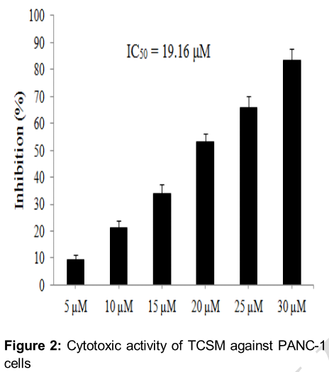
The cytotoxic activity of TCSM against PANC-1 cells was evaluated by MTT assay. PANC-1 cells were seeded on 96-well culture plates with DMEM medium. After 24 h of incubation, cells were treated with TCSM at concentrations of 0 (for control), 5, 10, 15, 20, 25 and 30 μM. After 48 h, MTT was added into every well, and cells were cultured at 37 °C in 5 % CO2 and 95 % O2 for another 3 h. Then the optical density (OD) was measured at 570 nm with a micro plate reader (Thermo, USA). The inhibition rate of TCSM against PANC-1 cells could be determined by the following equation: inhibition rate (%) = (OD control – OD treatment)/OD control × 100 %.
Flow cytometry analysis
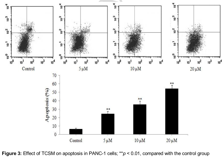
After treatment with TCSM at concentrations of 0 (for control), 5, 10, and 20 μM for 48 h, PANC-1 cells were collected and washed with phosphate buffered saline (PBS, 0.2 mol/L, pH = 7.0). Then the washed PANC-1 cells were re-suspended in staining buffer and stained with Annexin V-FITC/PI, and the PANC-1 cells were quantified by flow cytometry and analyzed by the Cell Quest Acquisition software (FACS Calibur, Becton Dickinson, USA).
Western blot analysis
After treatment with TCSM at concentrations of 0 (for control), 5, 10, and 20 μM for 48 h, total proteins of PANC-1 cells were extracted, and their concentration was determined by an Enhanced BCA Protein Assay Kit. Then, total protein (35 μg) were separated on 12 % SDS/PAGE and transferred onto a PVDF membrane. After blocking with 5 % fat-free milk, the membranes were incubated with primary antibodies including anti-β-actin, anti-Bcl-2, anti-Survivin, anti-Bax, anti-Smac, anti-cytochrome c, anti-c-caspase-3, anti-c-caspase-9 at 4 °C overnight. Membrane was washed with Tris buffered saline-Tween (TBS-T) and further incubated with HRP-conjugated secondary antibody for 1 h at room temperature in TBS-T. After another rinse, all proteins were detected by chemiluminescence. Additionally, β-actin was selected as an inner control to assess protein loading.
Tumor xenograft model
PANC-1 cells (2 × 10⁶ cells/nude mouse) were subcutaneously injected in the right flank of nude mice to establish the tumor xenograft model. When the tumor size grew to 2 - 3 mm in diameter, nude mice were randomly divided into control and treatment groups (n = 10) and given the following treatments: control group (0.5 % DMSO, intraperitoneal injection, ip) and treatment group (20 mg/kg TCSM, dissolved in 0.5 % DMSO, ip) once a day for 20 days. The body weight and tumor sizes (length and width) of nude mice were measured every 5 days with a vernier caliper, and the tumor volumes were calculated by the formula: tumor size (volume) = 0.52 × length (mm) × width2 (mm2) [10]. Finally, the tumor tissues of nude mice were removed, collected and used to determine the expressions of Bcl-2, Survivin, Bax, Smac, c-caspase-3, c-caspase-9 by western blot assay as described above.
Statistical analysis
All data are presented as mean ± standard deviation (SD). One-way ANOVA was used to analyze differences between control and treatment groups by Dunnett test with the aid of SPSS 21.0. Differences were recognized as statistically significant at p < 0.05.
Results
Identity and purity of TCSM
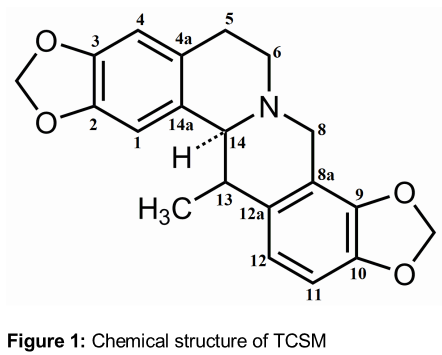
The results of mass spectrometry indicated that the molecular formula of target analyte was C20H19NO4. Comparing the NMR data of target analyte with existing literature [11], the target analyte, a colorless pinnate crystal, was identified as TCSM. 1H-NMR (400 MHz, CDCl3) δ: 6.54 (1H, s, H-1), 6.47 (1H, s, H-4), 2.48-3.20 (4H, m, H-5, H-6), 3.38 (1H, d, J = 15.2 Hz, Ha-8), 3.98 (1H, d, J = 15.2 Hz, Hb-8), 6.57 (1H, d, J = 8.1 Hz, H-11), 6.58 (1H, d, J = 8.1 Hz, H-12), 3.34 (1H, br. q, J = 6.9 Hz, H-13), 3.81 (1H, br. s, H-14), 5.90 and 5.98 (2H, d, J = 1.4 Hz, 2, 3 -OCH2O-), 5.96 and 6.05 (2H, d, J = 1.4 Hz, 9, 10 -OCH2O-), 1.04 (3H, d, J = 6.6 Hz, H-13-CH3); 13C-NMR (100 MHz, CDCl3) δ: 105.3 (C-1), 146.0 (C-2), 145.4 (C-3), 10.7.8 (C-4), 129.5 (C-4a), 29.4 (C-5), 50.8 (C-6), 52.9 (C-8), 116.3 (C-8a), 142.7 (C-9), 144.3 (C-10), 106.4 (C-11), 121.0 (C-12), 135.9 (C-12a), 38.4 (C-13), 63.4 (C-14), 129.7 (C-14a), 100.5 (2, 3 –OCH2O-), 100.9 (9, 10 –OCH2O-), 18.4 (13-CH3). The chemical structure of TCSM is shown in . The purity of TCSM was more than 95.3 %, which was determined by HPLC area normalization method.
Cytotoxic activity of TCSM against PANC-1 cells
The results of MTT assay indicated that the inhibition rates of TCSM in PANC-1 cells were between 9.28 and 83.23 %, and the IC50 value was 19.16 μM. Namely, TCSM showed good cytotoxic activity against PANC-1 cells. The results are shown in .
TCSM induces apoptosis in PANC-1 cells
Flow cytometry was used to study whether the cytotoxic activity of TCSM against PANC-1 cells was related to apoptosis. The results of flow cytometry suggested that the cytotoxic activity of TCSM was related to apoptosis. As shown in , the apoptosis rates were 6.12, 24.45, 35.26 and 54.16 % after treatment with TCSM at concentrations of 0 (for control), 5, 10 and 20 μM, and compared with the control group, the apoptosis rates of TCSM groups were significantly (p < 0.01) increased.
Effects of TCSM on the expressions of mitochondria-mediated apoptotic proteins
Western blot was used to study the pro-apoptotic mechanism of TCSM in PANC-1 cells. As shown in Figures 4 and 5, after PANC-1 cells were treated with TCSM at concentrations of 5, 10 and 20 μM, the expressions of anti-apoptotic proteins (Bcl-2 and Survivin) were significantly (p < 0.01) down-regulated, the expressions of pro-apoptotic proteins (Bax, Smac, c-caspase-3 and c-caspase-9) were significantly (p < 0.01) up-regulated, and the release of cytochrome c from the mitochondria to the cytoplasm was significantly (p < 0.01) promoted, compared with the control group.
Effects of TCSM on PANC-1 cell-induced xenograft model
PANC-1 cell-induced xenograft model was used to confirm whether TCSM could inhibit the growth of PANC-1 cell-induced tumor and regulate the expressions of apoptotic proteins in vivo. The results of xenograft assay indicated that TCSM (20 mg/kg once a day for 20 day) significantly (p < 0.05 or 0.01) inhibited the growth of PANC-1 cell-induced tumor without any effect on the body weight of the nude mice during the 20 days’ observation period, compared with the control group. Moreover, the results of western blot of tumor issues indicated that TCSM significantly (p < 0.01) down-regulated the expressions of anti-apoptotic proteins (Bcl-2 and Survivin) and up-regulated the expressions of pro-apoptotic proteins (Bax, Smac, c-caspase-3 and c-caspase-9) in vivo, compared with the control group. The results are shown in Figures 6 and 7.
Discussion
In the present study, we investigated the cytotoxic activity of TCSM against PANC-1 cells and possible mechanisms of action for the first time by MTT, flow cytometry, Western blot and xenograft assays.
MTT assay is a commonly used method to study the cytotoxic activity of compound against cells in vitro [12]. Flow cytometry is a commonly used method to investigate whether the cytotoxic activity of compound is related to apoptosis [13]. The results of MTT assay () indicated that TCSM showed good cytotoxic activity against PANC-1 cells with an IC50 of 19.16 μM. The results of flow cytometry assay () confirmed that the cytotoxic activity of TCSM against PANC-1 cells was related to apoptosis.
Western blot is a commonly used method to explore anticancer mechanism at the protein level, and the effect of compound on tumor growth and body weight of nude mice are two important indices in vivo [14,15]. Proteins including Bcl-2, Survivin, Bax, Smac, cytochrome c, c-caspase-3 and c-caspase-9 are very important markers in mitochondria-mediated apoptotic pathway [16]. The relations among these apoptotic proteins are rather complex and described as follows [17-21]. When cells suffer from apoptotic stimuli, pro-apoptotic proteins (Smac and cytochrome c) are released from the mitochondria to the cytoplasm, but their release can be inhibited by anti-apoptotic protein (Bcl-2). However, the function of Bcl-2 can be inhibited by pro-apoptotic protein (Bax).
Cytochrome c along with apoptotic protease activating factor-1 (Apaf-1), caspase-9 and ATP can form apoptosome in the cytoplasm to activate caspase-9 (c-caspase-9), and then c-caspase-9 can promote the activation of caspase-3 (c-caspase-3), which can lead to apoptosis of cells. The results of western blot assay (Figures 4 and 5) suggested that TCSM down-regulated the expressions of anti-apoptotic proteins (Bcl-2 and Survivin), up-regulated the expressions of pro-apoptotic proteins (Bax, Smac, c-caspase-3 and c-caspase-9) and promoted the release of cytochrome c from the mitochondria to the cytoplasm in PANC-1 cells.
The results of the xenograft assay (Figures 6 and 7) in nude mice indicated that TCSM inhibited the growth of PANC-1 cell-induced tumor without any effect on the body weight of the nude mice by regulating the expressions of mitochondria-mediated apoptotic proteins in vivo as described above.
Conclusion
TCSM exhibits cytotoxic activity against PANC-1 cells in vitro and in vivo through mitochondria-mediated apoptotic pathway. Further investigation into the mechanism of action of TCSM is required.
References
Archives


News Updates